
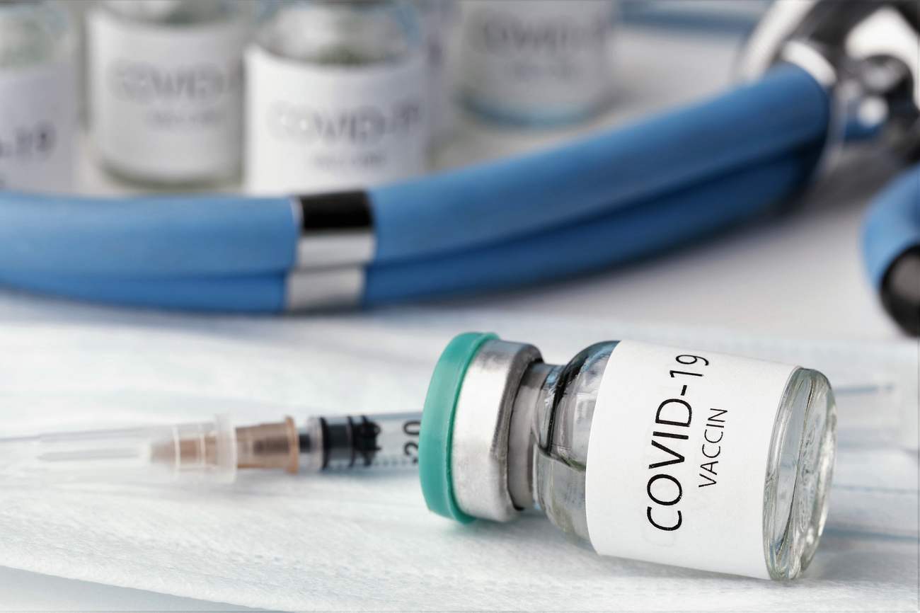
SARS-CoV-2-Induced Neurological Disorders in Symptomatic Covid-19 and Long Covid Patients: Key Role of Brain Renin-Angiotensin System

Par Jean-Marc Sabatier*
et Ziad Fajloun*
Covid-19 caused by the SARS-CoV-2 virus generates over a hundred symptoms and illnesses [1]. After five weeks, about 20-30% of Covid-19-affected individuals will still be experiencing one or more of the initial disease symptoms (about 10% will be affected six months later) [2]. To date, more than 54 million people worldwide are suffering from the long-term effects of Covid-19 diseases. Long-term symptoms and/or illnesses associated with Covid-19 are various on organs, tissues, and brain. In brain, they varied from severe incapacitating exhaustion to neurocognitive issues, neurophysiological disturbances (dizziness, memory loss, spatial disorientation, headache), and loss of smell (anosmia) and/or taste (ageusia). These more or less severe adverse effects may be temporary and cyclical in character (they may emerge, vanish, and then reappear) [1]. The “Long Covid” term refers to these post-Covid-19 consequences. The World Health Organization (WHO) defines Long Covid as a condition that lasts more than 2 months and cannot be explained by another diagnosis [3].
A second pathway
Various explanations have been proposed as to the cause of Long Covid in terms of pathophysiology. The first is the persistence of a dormant SARS-CoV-2 virus in the body that has not been entirely eradicated (chronic viral infection with transient reactivations), which has harmful effects [4]. A second pathway is SARS-CoV-2-induced hyperinflammation, which would result in the reactivation of endogenous microorganisms (for example, Epstein-Barr virus, a member of the herpes family found in 95% of the world’s adult population) and have a deleterious influence on the organism [5]. The presence of autoimmune antibodies directed against one or more host proteins (such as coagulation factor VIII, the PF4 protein of blood platelets, and others) is the third possibility [6].
A physiological hormonal system
Such autoimmune antibodies are thought to alter the normal functioning of the body in symptomatic individuals. As observed in infected patients, autoimmune antibodies may also be directed against components of the renin-angiotensin system (RAS), a physiological hormonal system that controls renal, pulmonary, and cardiovascular autonomic functions, as well as innate immunity and the gut flora (and likely the mouth flora). These autoimmune antibodies include anti-ACE2 receptor (angiotensin-2 converting enzyme receptor) antibodies, which typically binds and degrades angiotensin-2 but also serves as a binding site for SARS-CoV-2, interfering thus with hormone degradation. They also include anti-angiotensin-2 antibodies and anti-AT1R (angiotensin-2 type 1 receptor) antibodies, noting that excess angiotensin-2 overactivates the AT1R which is directly accountable for Covid-19 pathologies [7, 8]. Such autoimmune antibodies have the potential to impair metabolic pathways as well as physiological processes that are crucial in organ and tissue functions. As a result, these antibodies would be responsible for the damaging effects caused by such disruptions or dysfunctions [6]. The other pathophysiological tracks for Long Covid would be initial organic lesions whose persistent symptoms are consequences (epithelial dysfunction, pulmonary fibrosis, cerebral microglia, etc.), as well as persistent abnormal immune and inflammatory reactions which become complicated (microcirculation, coagulation, fibrosis, neuro-inflammation, auto-immunity, …)
Disruptions or dysfunctions
Concerning the abnormalities linked with Long Covid in some patients, we contend that these dysfunctions might be due to the persistence of the virus which has not been completely eradicated, at the systemic, tissue, and/or cerebral levels [4]. SARS-CoV-2 would be directly involved in the development of neurological disorders such as vertigo, memory loss, spatial disorganization, headache, meningitis, cerebral thrombosis, ischemic stroke, delirium, dementia, and other nervous pathologies by disrupting the brain (central nervous system (CNS)) renin-angiotensin system (called RAS-b for the brain). SARS-CoV-2 is able to infect brain cells – neurons, astrocytes, microglial cells and probably oligodendrocytes – by binding to the ACE2 receptor in these cells.
Brain damage
Theoretically, the possible entry routes of SARS-CoV-2 into the CNS are (i) via the disruption of the blood-brain barrier (ii) the blood-barrier cerebrospinal fluid, (iii) the trans-synaptic viral diffusion, and (iv) via the circumventricular organs. After entering the ENT sphere, SARS-CoV-2 may assault the olfactory bulb on the floor of the skull, passing through the olfactory epithelium in the upper nasal cavity, which is located nearby (below the olfactory bulb). This explains the phenomenon of anosmia, but also ageusia, since this same olfactory bulb also manages information concerning taste [9].
SARS-CoV-2-induced brain damage is revealed by imaging in Long Covid patients [10]. Some areas of the brain appear to have been harmed, while others remain undamaged. Cerebral hypoactivity is found in the altered brain areas, resulting in a decrease in glucose intake. We assume that a dysfunctioning of RAS-b in the CNS is responsible for these alterations. The loss of peripheral gray matter in both cerebral hemispheres reflects the damage to nerve cells induced by the viral infection (cerebral cortex made up of neuronal bodies) [11].
The blood-brain barrier
In the CNS, the Spike protein of SARS-CoV-2 binds to the ACE2 of nerve cells and interferes with the degradation of angiotensin-2. This leads to an excess angiotensin-2 which overactivates the RAS-b AT1R [12,13]. RAS-b dysfunction causes multiple deleterious effects, including vasoconstriction (decrease in blood vessel diameter thereby reducing cerebral microcirculation), inflammation, and oxidative stress. The blood-brain barrier governs the flow of nutrients to the brain, among other functions, and a diminished cerebral irrigation could have a deleterious impact on brain function, causing it to move into a protective or standby state [12]. It is also plausible that pro-inflammatory events (such as pro-inflammatory cytokines released by microglial cells that operate as immune system macrophages in the brain), oxidative stress, and/or glucose intolerance caused by AT1R overactivation, are to blame for the observed brain hypoactivity.
Different functional properties
The observed disparities in altered brain areas can be explained by the fact that RAS-b is significantly expressed differently in various brain regions [14]. In “unaffected” brain areas, the AT1R (the preferred target of angiotensin-2) is undetectable (by the experimental techniques used) or strongly under-represented compared to AT2R (angiotensin-2 type 2 receptor), another potential target of angiotensin-2, but of low affinity. As a result, with the absence or under-representation of AT1R, angiotensin-2 (hormone and neuropeptide) will bind primarily to the AT2R, which has protective and reparative actions (vasodilation, axonal regeneration, and neuronal development) that are diametrically opposed to AT1R. Thus, the AT2R promotes neuronal survival and protects against brain damage [15]. Moreover, it has been shown that the AT1R can be over-expressed in case of cell activation. The AT1R can also function as a heterodimer that is can associate with an AT2R or MasR (Mas receptor), or others like AT4R (angiotensin-2 type 4 receptor) or MrgD (G-protein-coupled receptor for angiotensin (1-7)). The hybrid dimeric receptor (AT1R/AT2R or AT1R/MasR) formed has different functional properties. It is worth mentioning that at the level of the brain RAS-b, there is also pro-renin, which binds to the pro-renin receptor of nerve cells and has deleterious effects similar to those caused by the AT1R when the latter is overactivated. In fact, although the properties of this receptor are still poorly characterized, it appears that activation of the pro-renin receptor leads to an overactivation of the RAS-b [16,17].
Remarkably, there is another intracrine RAS-b located inside nerve cells and localized on the membranes of the cell nucleus, the endoplasmic reticulum, the secretory vesicles, and the mitochondria (the cell energy centers) in the CNS. This intracrine RAS-b is “autonomous” in comparison to the systemic endocrine RAS, which drives the various organs and tissues of the body through endocrine, autocrine, and paracrine actions. The RAS-b also controls the release of neurotransmitters (acetylcholine, dopamine, vasopressin, and others) and the production of NO (nitric oxide) which directly affects brain activity (actions on inflammation, immunity and memory phenomena) [14].
The brain needs vitamin D
Vitamin D is a molecule that should be prioritized for overcoming the detrimental effects of the virus (particularly in the brain), both in the prophylaxis and in the treatment of Covid-19 and Long Covid [18-20]. Vitamin D can affect directly or indirectly pro-renin and pro-renin receptor activation because it lowers renin synthesis and so has a favorable regulatory (negative regulatory) effect on the RAS [19]. Thus, in concert with reduction of systemic RAS overactivation, vitamin D -specifically D3/cholecalciferol- may successfully prevent and/or reverse brain damage in SARS-CoV-2 infected persons with neurological disorders connected to dysfunctional RAS-b.
The regions of the brain whose cells are rich in vitamin D (calcitriol) receptors are the cerebral cortex and the hippocampus. The cerebral cortex corresponds to the peripheral gray matter that covers our two cerebral hemispheres. The cerebral cortex is the seat of intelligence, voluntary movement, consciousness, sensitivity…It plays a key role in motor skills, sensitivity, sensoriality (ability to perceive sensations for a living being) or sensoricity (all of the sensory organization of the human body). The cortex has an indispensable role in language and memory.
Some people (previously infected or uninfected with SARS-CoV-2) have been reported to suffer from Long Covid after SARS-CoV-2 vaccination. Besides the purely psychological aspects in some of these people, it appears possible that the vaccine Spike protein is capable of causing the RAS-b to malfunction in some cases, just as it is capable of causing the endocrine systemic RAS (which acts on the various organs and body tissues) to malfunction.
*Jean-Marc Sabatier, research director at the CNRS and doctor in Cell Biology and Microbiology, affiliated with the Institute of Neuro Physiopathology (INP), at the University of Aix-Marseille.
* Ziad Fajloun is a University Professor at the Faculty of Science &EDST Lebanese University Tripoli. Lebanon.
Références
[1] Hofer U. Dose-dependent COVID-19 symptoms. Nat Rev Microbiol 2021; 19: 682. Doi.org/10.1038/s41579-021-00634-4
[2] Mandal S, Barnett J, Brill SE, Brown JS, Denneny EK, Hare SS, Heightman M, Hillman TE, Jacob J, Jarvis HC, Lipman MCI, Naidu SB, Nair A, Porter JC, Tomlinson GS, Hurst JR. ‘Long-COVID’: a cross-sectional study of persisting symptoms, biomarker and imaging abnormalities following hospitalisation for COVID-19. Thorax. 2021; 76:396-398. Doi: 10.1136/thoraxjnl-2020-215818.
[3] NICE defines ‘long Covid’ as symptoms lasting more than 12 weeks. GEMMA MITCHELL, 30 Octobre, 2020; https://www.nursingtimes.net/news/coronavirus/nice-defines-long-covid-as-symptoms-lasting-more-than-12-weeks-30-10-2020/
[4] Kang H, Wang Y, Tong Z, Liu X. Retest positive for SARS-CoV-2 RNA of “recovered” patients with COVID-19: Persistence, sampling issues, or re-infection? J Med Virol. 2020; 92:2263-2265. doi: 10.1002/jmv.26114.
[5] Naendrup JH, Garcia Borrega J, Eichenauer DA, Shimabukuro-Vornhagen A, Kochanek M, Böll B. Reactivation of EBV and CMV in Severe COVID-19-Epiphenomena or Trigger of Hyperinflammation in Need of Treatment? A Large Case Series of Critically ill Patients. J Intensive Care Med. 2021; 18:8850666211053990. Doi: 10.1177/08850666211053990.
[6] Sacchi MC, Tamiazzo S, Stobbione P, Agatea L, De Gaspari P, Stecca A, Lauritano EC, Roveta A, Tozzoli R, Guaschino R, Bonometti R. SARS-CoV-2 infection as a trigger of autoimmune response. Clin Transl Sci. 2021; 14:898-907. Doi: 10.1111/cts.12953.
[7] Amiral J, Vissac AM, Seghatchian J. Covid-19, induced activation of hemostasis, and immune reactions: Can an auto-immune reaction contribute to the delayed severe complications observed in some patients? Transfus Apher Sci. 2020; 59:102804. Doi: 10.1016/j.transci.2020.102804.
[8] Lee DH, Heidecke H, Schröder A, Paul F, Wachter R, Hoffmann R, Ellrichmann G, Dragun D, Waschbisch A, Stegbauer J, Klotz P, Gold R, Dechend R, Müller DN, Saft C, Linker RA. Increase of angiotensin II type 1 receptor auto-antibodies in Huntington’s disease. Mol Neurodegener. 2014; 9:49.
Doi: 10.1186/1750-1326-9-49.
[9] Burks SM, Rosas-Hernandez H, Alejandro Ramirez-Lee M, Cuevas E, Talpos JC. Can SARS-CoV-2 infect the central nervous system via the olfactory bulb or the blood-brain barrier? Brain Behav Immun. 2021; 95:7-14. Doi: 10.1016/j.bbi.2020.12.031.
[10] Guedj E, Campion JY, Dudouet P, Kaphan E, Bregeon F, Tissot-Dupont H, Guis S, Barthelemy F, Habert P, Ceccaldi M, Million M, Raoult D, Cammilleri S, Eldin C. 18F-FDG brain PET hypometabolism in patients with long COVID. Eur J Nucl Med Mol Imaging. 2021; 9:2823-2833. Doi: 10.1007/s00259-021-05215-4.
[11] Douaud G, Lee S, Alfaro-Almagro F, Arthofer C, Wang C, McCarthy P, Lange F, Andersson JLR, Griffanti L, Duff E, Jbabdi S, Taschler B, Keating P, Winkler AM, Collins R, Matthews PM, Allen N, Miller KL, Nichols TE, Smith SM. SARS-CoV-2 is associated with changes in brain structure in UK Biobank. Nature 2022; 2. Doi: 10.1101/2021.06.11.21258690.
[12] El-Arif G, Farhat A, Khazaal S, Annweiler C, Kovacic H, Wu Y, Cao Z, Fajloun Z, Abi Khattar Z, Sabatier J.M. The renin-angiotensin system: a key role in SARS-CoV-2-induced COVID-19.
Molecules. 2021; 26: 6945. Doi: 10.3390/molecules26226945.
[13] Annweiler C, Cao Z, Wu Y, Faucon E, Mouhat S, Kovacic H, Sabatier J.M. Counter-regulatory ‘renin-angiotensin’ system-based candidate drugs to treat COVID-19 diseases in SARS-CoV-2-infected patients. Infectious Disorders-Drug Targets. 2020; 20: 407-408.
Doi: 10.2174/1871526520666200518073329
[14] Cosarderelioglu C, Nidadavolu L, George C, Oh2 ES, Bennett DA, Walston JD. and. Abadir PM. Brain Renin–Angiotensin System at the Intersect of Physical and Cognitive Frailty. Front. Neurosci. 2020; Doi.org/10.3389/fnins.2020.586314
[15] Annweiler C, Bourgeais A, Faucon E, Cao Z, Wu Y, Sabatier J.M. Neurological, cognitive and behavioral disorders during COVID-19: the nitric oxide track. Journal of American Geriatrics Society. 2020, 10.1111/jgs.166671. Doi: 10.1111/jgs.166671
[16] Alinaghi SA, Mehrtak M, Mohssenipour M, Mirzapour P, Barezegary A, Habibi P, Moradmand-Badie B, Afsahi AM, Karimi A, Heydari M, Mehraeen E, Dadras O, Sabatier JM, Voltarelli F. Genetic susceptibility of Covid-19: a systematic review of current evidence. Eur. J. Med. Res. 2021; 26: 46.
Doi: 10.1186/s40001-021-00516-8.
[17] Annweiler C, Cao Z, Papon N, Kovacic H, Sabatier J.M. Counter-regulatory renin-angiotensin system: an important line of research to understand and limit the severity of COVID-19. Infectious Disorders-Drug Targets. 2021; 21. Doi: 10.2174/1871526521999210827142839
[18] Annweiler C, Cao Z, Sabatier JM. Point of view: Should COVID-19 patients be supplemented with vitamin D? Maturitas. 2020; 140: 24-26. Doi.org/10.1016/j.maturitas.2020.06.003
[19] Annweiler C, Hanotte B, de l’Eprevier CG, Sabatier JM, Lafaie L, Célarier T. Vitamin D and survival in COVID-19 patients. A quasi-experimental study. J. Steroid Biochem. Mol. Biol. 2020; 204:105771. Doi: 10.1016/j.jsbmb.2020.105771.
[20] Cao Z, Wu Y, Faucon E, Sabatier J.M. SARS-CoV-2 & Covid-19: Key-roles of the renin-angiotensin system / Vitamin D impacting drug and vaccine developments. Infectious Disorders-Drug Targets, 2020; 20, 348-349 Doi: 10.2174/1871526520999200505174704